 loading
Light & VeritySkin-deep success storyA trio of students wins a $100,000 prize for a medical device. 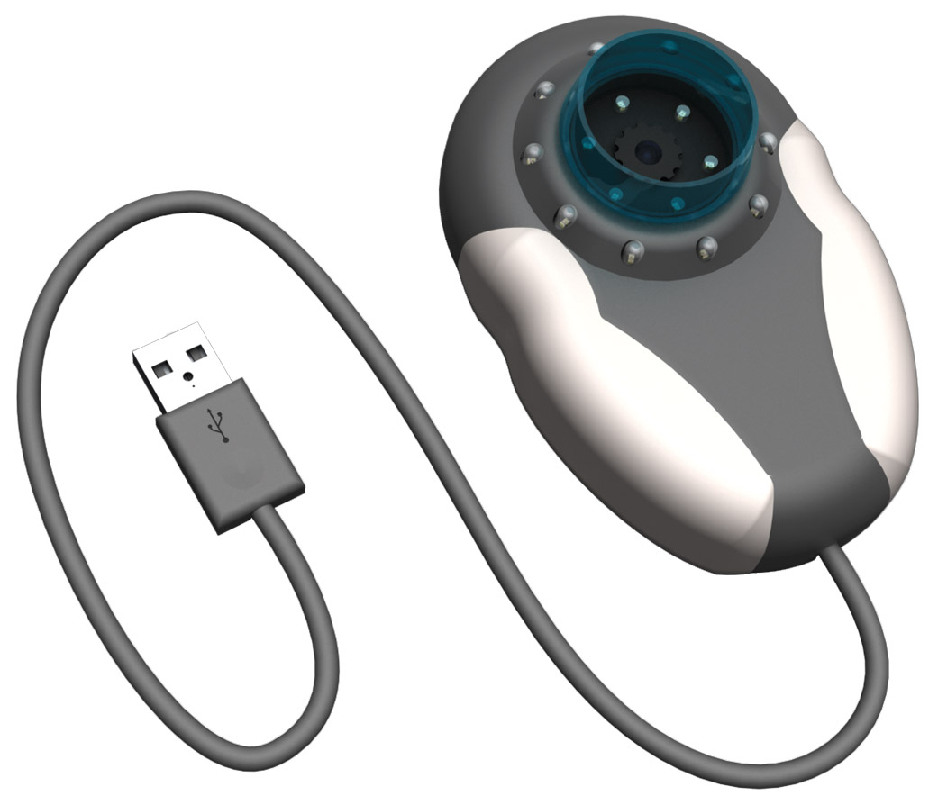 Courtesy Nickolas P. DemasUndergraduate engineering students came up with a 3-D camera to help doctors assess moles and lesions remotely. View full image
Intense late-night conversations in the dorm, burning the midnight oil to make a deadline, and living on a tight budget are everyday features of undergraduate life. But for three Morse College engineering majors, they were also part of the road to a $100,000 prize for an innovative new medical device. Juniors Elizabeth Asai, Elliot Swart, and Nickolas Demas won the cash prize from the Center for Integration of Medicine and Innovative Technology in June. Working 15-hour days to meet the competition deadline and keeping the prototype cost to $100, they developed a product they call a Stereoscopic Plug-and-Play Dermatoscope and Web Interface. It takes pictures of moles and other suspicious lesions to help patients and dermatologists catch melanomas earlier than existing methods. “We were competing with graduate students and PhD candidates, so we really didn’t know what to expect,” says Asai. Their approach was to take an existing piece of medical technology and literally add another dimension, enhancing a traditional dermatoscope to take three-dimensional images. “Elevation is one of the classic changes doctors look for, so just looking at a two-dimensional image isn’t helpful,” says Asai. The other innovation the students came up with was an interface for uploading the images onto a server, so doctors can study them remotely—making it easier to “monitor trends and changes over time and catch a lesion going haywire much earlier,” says Demas. They expect that the product would cost about $35 to mass-produce. After creating a working prototype, the group put their product through clinical trials. Working with four dermatologists from Yale and 50 patients, the students captured images of the patients’ lesions using their device. Each image was reviewed by an off-site physician. The same lesion was then examined in person by a different physician. The results: there was not a single instance, says Demas, in which the on-site doctor recommended a biopsy but the off-site doctor did not. The students plan to use their prize money to buy a three-dimensional printer and prepare another clinical study. “We literally went from an idea on paper to a fully tested prototype in three and a half months,” said Demas. “There are a number of improvements we expect to make to bring our product to the next level.”
The comment period has expired.
|
|
 Print
Print Email
Email