 loading
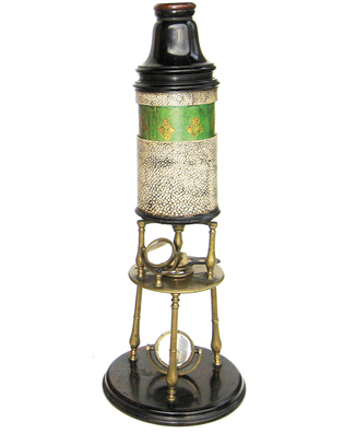
Arts & CultureUp closeObject lesson: a very short history of the microscope. Thomas L. Lentz ’64MD is a Yale senior research scientist and professor emeritus of cell biology, and a curatorial affiliate at the Peabody Museum.  Yale Peabody MuseumA tripod microscope made by Edmund Culpeper in London, about 1725. This instrument and many others, illustrating the entire history of microscopy, were donated to the Yale Peabody Museum by the author. To see more microscopes from the collection, go to peabody.yale.edu/exhibits/curators-choice/microcosm-wonders. View full imageIt all started with spectacles. The first pairs of spectacles were made in Italy at the end of the thirteenth century—showing that people knew about the magnifying properties of glass lenses even in that early period. It seems likely that spectacle makers experimenting with multiple lenses invented both the telescope and the compound microscope, as they both consist of two or more lenses held in a tube. But the first microscope we know of did not show up until centuries later: credit is usually given to Hans Jansen and his son Zacharias, spectacle makers in Holland, who built their instrument around the year 1595. The microscope shown here was made in about 1725, in London, by Edmund Culpeper. This particular instrument stands 14 inches high and is crafted of lignum vitae, boxwood, brass, pasteboard, green vellum with gold tooling, and shagreen—the skin of a ray. Like most of the microscopes that came before it, it takes the form of a cylindrical tube with an eye lens at one end and an objective lens at the other and supported by a small tripod. (The objective lens produces a magnified image of the specimen being examined; this image is further magnified by the eye lens.) But Culpeper devised two improvements on the earlier instruments, improvements still used in light microscopes today. He raised the stage, which holds the specimen, above table level, making it more accessible; and he inserted a concave mirror below the stage to illuminate the specimen. (Yale also has another Culpeper-type microscope, purchased by Yale College in 1734. That instrument, made by John Loft after Culpeper’s design, was the first compound microscope in America.) Several scientific discoveries were made using very early microscopes. In 1661, Marcello Malpighi discovered capillaries in the lung—thereby proving William Harvey’s theory of the continuous circulation of blood. Other important observations were made by Robert Hooke (1635–1703), who was the first to use the word “cell.” Antonie van Leeuwenhoek (1632–1723) made the first observations of protozoa, bacteria, spermatozoa, cardiac muscle, and the banded pattern of muscle fibers. Nevertheless, until the 1800s microscopes mostly served as sources of entertainment and amusement for the upper classes. Their objective lenses suffered from low resolution and flaws that distorted the images, so they were of limited use for scientific investigation. But the first half of the nineteenth century saw rapid improvements in microscope design and function. Around 1840 in London, instrument maker Andrew Ross and optics theoretician Joseph J. Lister perfected lenses that were no longer plagued by optical error—transforming the microscope into an important scientific tool. Further improvements were made in the late 1800s. Although the twentieth century would see many additional advances in light microscope design, by the end of the nineteenth century the instrument had been perfected to the point where it achieved its theoretical limit of resolution of 0.2 microns.
The comment period has expired.
|
|